Team:DTU-Denmark/Project/Chip

Lab-on-a-disc Screening
To finalize the Synthesizer project we thought about a screening method for our MAGE method to distinguish bacterial colonies producing non-ribosomal peptides (NRPs) of interest. A few NRPs we worked with, such as tyrothricin, tyrocidine and surfactins distinguish cytotoxic properties towards red blood cells [1][2][3]. This fact inspired us to use it as a screening method for certain bacterial products in our MAGE method.
Achievements
- UV-vis absorbance spectra prove red blood cells are lysed in presence of certain NRPs. The experiments should be repeated with a use of fresh blood cells.
- Proof of a concept of a lab-on-a-disc.
Background
Certain groups of non-ribosomal peptides (NRPs), including commonly used antibiotics, display non-specific cytotoxicity. [1][2][3] Therefore, the substances are commonly used on external surfaces, topically[4], as intravascular injection causes hemolysis of red blood cells. It has also been proved that certain concentrations of these products kill animal models.[1] This supports our screening concept by distinguishing the presence of specific antibiotics in bacterial cultures by red blood cell lysis.
UV- vis absorbance spectrum depicts the amount of light absorbed by a measured solution or compounds included in a sample. Moreover, the absorbance spectra within a range of 300-800nm show changes depending on the state of the red blood cells. [5][6]
Methods
To study cell lysis, the absorbance of fresh blood cells spiking with non-ribosomal peptides was measured. To perform the experiments, we measured the UV-vis absorption spectra by DS-11 + Spectrophotometer (DeNovix) to detect red blood cell lysis. The measurements were performed in cuvettes by spectrophotometer and by nanodrop device. In this subproject three experiments were investigated. All of them were investigating UV-vis absorption spectra of red blood cells in the presence of different concentration of NRPs. The experiments focused on investigation of anticoagulants in blood samples, cytotoxic properties of tyrothricin and sensitivity of red blood cells in presence of unknown concentrations of Tyrocidine produced by Bacilus brevis. Absorbance spectra of blood samples in different conditions were recorded between 300-800nm. Further description is found in Lab Notebook.
Results
The UV-vis absorption spectra of red blood cells suspended in the absence or presence of antibiotic (Figure 1) proves toxicity of tyrothrycin towards blood cells. The drop of the green curve around 600nm indicates less absorbance of red blood cells in the sample; at some point the curve reaches the “0” level indicating cell lysis.

Figure 1. Comparison of blood cells in different buffers.
grey line: blood cells anticoagulated in heparin with 0.15M NaCl; non-lysed
green line: blood cells anticoagulated in heparin with unknown concentration of Tyrothrycin; lysed
Decrease of UV absorbance around 600nm occurred in all of the blood samples with tyrothricin (Figure 2.) indicating cell lysis.

Figure 2. Comparison of blood cells in presence of antibiotic over time.
pink and black line : unknown concentration of Tyrothrycin measured after 30sec with blood cells anticoagulated in heparin; green line: unknown concentration of Tyrothrycin measured after 120sec with blood cells anticoagulated in heparin.
The same concept of experiment was performed with specified concentrations of tyrothricin, measured by a nanodrop device at different time points.
Figure 3. Comparison of blood cells (anticoagulated with heparin) in presence of 0,15M NaCl and different concentrations of tyrothricin after 45-60sec of incubation. Nanodrop measurement.
The absorbance of spectra in all of the cases (see Figure 3) shows cell lysis, but not full cell lysis. It might occurred due to a short time of incubation of the samples as the measurement was done only after 45-60 sec. However, in this experiment blood sample resuspended in 0,15M NaCl should not lyse the red blood cells, as sodium chloride does not have properties that burst cells by osmotic pressure. Despite this, the sample (red curve) still shows some turbidity around 600-700nm in comparison to other measurements. The part-cell lysis could happen due either to a use of a narrow orifice of pipette tip that bursts the cells (mechanical forces) or due to hemolysis of the cells, as on the day of this measurement the blood samples were three days old.[6] We can conclude that the antibiotic lyses the red blood cells but we cannot determine its specificity very accurately.
The Figure 4 below, depicts also UV-vis absorbance spectra of non-ribosomal peptides delivered from Bacillus brevis colonies. The samples were previously detected by a LCMS and it was proved they contained tyrocidine.
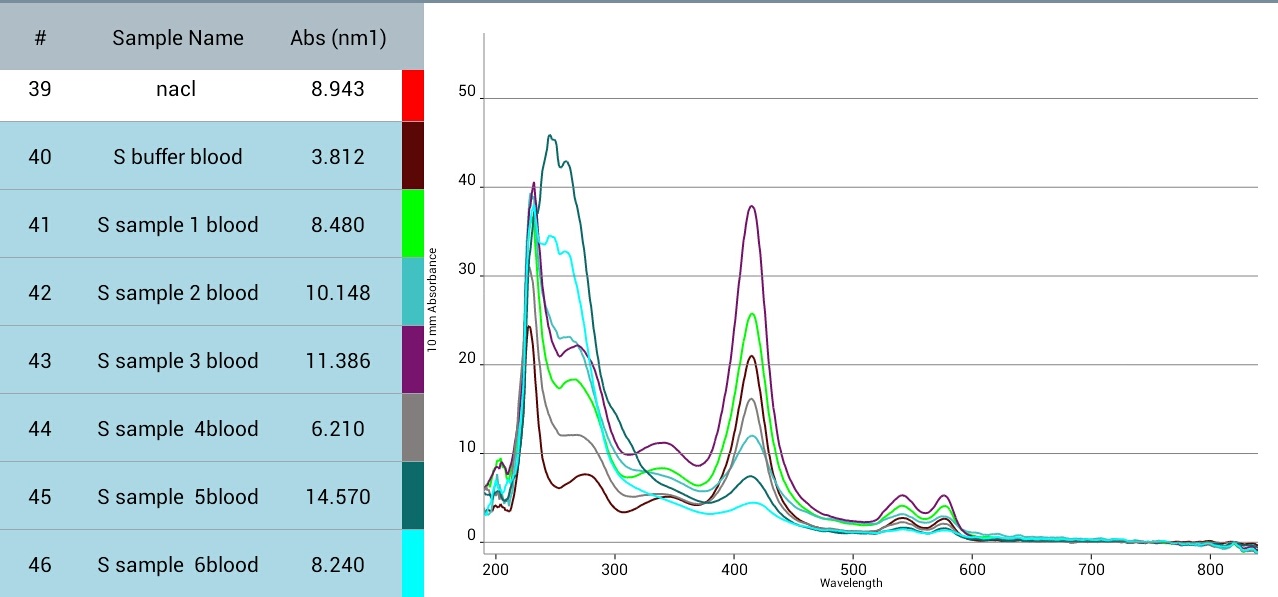
Figure 4. UV-vis absorption spectra of blood anticoagulated with heparin in presence of unknown concentrations of tyrocidine after 45-60sec of incubation. Nanodrop measurement.
In this experiment cell lysis is also observed in all samples. The control sample should retain blood cell stability, however it did not. It may be due to the fact the blood cells were three days old, as in the previous experiment or they were destroyed due to mechanical forces while pipetting their volumes on nanodrop. Preferably, the experiments should be repeated. However, the high peaks of a few samples might suggest complete cell lyses due to the presence of antibiotic.
Discussion
Even though the results are not 100% precise they are a basic and positive foundation for our idea of the screening method. Additionally, current knowledge about the properties of these peptides gives strong support to the theory that the lab-on-a-disc screening of NRP products of bacterial transformants for the Synthesizer project is very likely to be viable and has the potential to be a low-cost, quick and simple way of detection and screening of interesting transformants.
Concept of lab-on-a-disc
The rotating disc would be an easy and low-cost way to screen specificity of bacterial colonies. Our inspiration came from the start-up company BluSense Diagnostics, who specialise within microfluidics and rotor devices.
As we mentioned above, our idea for a selection of bacterial colonies of interest is based on the toxicity of several MAGE products such as tyrothricin, surfactins or tyrocidine towards red blood cells [1][2][3][5] . The rotor disc would enable visual screening (detection) of bacterial colonies by mixing both small volumes of red blood cells and supernatants of bacterial culture. As mentioned before, red blood cells in the presence of specific NRPs are lysed. This aids in the selection of the transformants of interest. The primary idea of the disc would enable us to screen five different colonies at the same time. Figure 5., below, displays a simple set-up of lab-on-a-disc device.
Figure 5. Illustration of lab-on-a-chip concept for screening the MAGE products.
The use of the disc for observing cell lysis is very easy (Figure 5.). According to methods used in BluSense Diagnostics, it consists of a few steps. First, loading the bacterial supernatant and blood volume to specific inlets (Figure 6.). Then the disc starts to rotate. At the same time the bacterial supernatant is moved to the mixing/detection chamber while the certain volume of the blood sample is measured off in a specific chamber and with the next rotation the specified volume gets to the mixing chamber with the bacterial supernatant (see picture A, Figure 7.). Next, the disc stops rotating, before again beginning to accelerate and decelerate for a certain time period to mix the fluid (see picture B, Figure 7.). The disc stops and after a time period, the blood cells are either lysed or not, depending on the presence of specific antibiotics.

Figure 6. Visual presentation of an original disc design. Disc provided by BluSense Diagnostics.
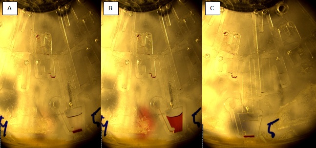
Figure 7. Visualisation of rotations influencing samples’ microflow.
A)Two different solutions are transferred to the mixing chamber. B) Mixture after several rotations of the disc. C) Visible cell lysis.
Pictures provided by BluSense Diagnostics.
The idea behind this concept still needs investigation in terms of the sensitivity of the test, the interaction between bacterial supernatant-red blood cells, the most efficient method of extraction of NRPs for screening, optimization of mixing and microflow within the disc. However, it is very likely that this design would be a postive screening method for MAGE.
References
- Dubos, R. J. (1941). THE PRODUCTION OF BACTERICIDAL SUBSTANCES BY AEROBIC SPORULATING BACILLI. Journal of Experimental Medicine, 73(5), 629–640. doi:10.1084/jem.73.5.629
- Findlay R. D., Taeusch H. W., David-Cu R., Walther F. J., Pediatr Res., Lysis of red blood cells and alveolar epithelial toxicity by therapeutic pulmonary surfactants.(1995); 37(1):26-30.
- Pape W. J., Pfannenbecker U., Hoppe U. (1987-1988 Fall) Validation of the red blood cell test system as in vitro assay for the rapid screening of irritation potential of surfactants.Mol Toxicol.;1(4):525-36
- https://pubchem.ncbi.nlm.nih.gov/compound/tyrothricin#section=Top
- Jiang, N., Tan, N. S., Ho, B., & Ding, J. L. (2007). Measurement of the red blood cell lysis by bacterial hemolysin. Protocol Exchange. doi:10.1038/nprot.2007.483
- Jávorfi T., Erostyák J., Gál J., Buzády A., Menczel L., Garab G., Razi Naqvi K. J. Photochem Photobiol B. (2006) Quantitative spectrophotometry using integrating cavities. 82(2):127-31. Epub 2005 Nov

Department of Systems Biology
Søltofts Plads 221
2800 Kgs. Lyngby
Denmark
P: +45 45 25 25 25
M: dtu-igem-2015@googlegroups.com