Difference between revisions of "Team:Oxford/Design"
Weikongquee (Talk | contribs) |
|||
| (31 intermediate revisions by 4 users not shown) | |||
| Line 2: | Line 2: | ||
<html> | <html> | ||
| − | < | + | <style> |
| − | </ | + | /*main colour*/ |
| + | .navbar-default .navbar-brand, | ||
| + | .contents-sidebar .nav>.active>a, | ||
| + | .contents-sidebar .nav>.active>a, | ||
| + | .contents-sidebar .nav>li>a:hover, | ||
| + | .contents-sidebar .nav>li>a:focus, | ||
| + | h2, | ||
| + | h1 { | ||
| + | color: #723EC3; | ||
| + | } | ||
| + | .contents-sidebar .nav>.active>a, | ||
| + | .contents-sidebar .nav>li>a:hover, | ||
| + | .contents-sidebar .nav>li>a:focus { | ||
| + | border-left: 2px solid #723EC3; | ||
| + | } | ||
| + | .image.lightbox, #notebook-key-button { | ||
| + | background-color: #723EC3; | ||
| + | } | ||
| + | |||
| + | /*complimentary colour*/ | ||
| + | .navbar-default .navbar-brand:hover, | ||
| + | .definition:hover, .definition:focus, | ||
| + | ol li::before, | ||
| + | .slim ul li:before, | ||
| + | .table>thead>tr>th, | ||
| + | .algorithm ol li::before, | ||
| + | .quote, | ||
| + | .quote h3 { | ||
| + | color: #B131BE; | ||
| + | } | ||
| + | .definition { | ||
| + | border-bottom: 1px dotted #B131BE; | ||
| + | } | ||
| + | .popover-title { | ||
| + | background-color: #B131BE; | ||
| + | } | ||
| + | .popover.right>.arrow::after{ | ||
| + | border-right-color: #B131BE; | ||
| + | } | ||
| + | .popover.bottom>.arrow::after { | ||
| + | border-bottom-color: #B131BE; | ||
| + | } | ||
| + | .popover.left>.arrow::after { | ||
| + | border-left-color: #B131BE; | ||
| + | } | ||
| + | </style> | ||
<body> | <body> | ||
| − | <div class="container-fluid page-heading" style="background-image: url(https://static.igem.org/mediawiki/2015/ | + | <div class="container-fluid page-heading" style="background-image: url(https://static.igem.org/mediawiki/2015/1/10/Ox_DesignHeader.jpeg)"> |
<h3>Design</h3> | <h3>Design</h3> | ||
</div> | </div> | ||
| Line 12: | Line 57: | ||
<div class="col-md-9"> | <div class="col-md-9"> | ||
<div class="slim"> | <div class="slim"> | ||
| + | <div class="section" id="preface"> | ||
| + | <h2>Preface</h2> | ||
| + | <p> | ||
| + | This page comprehensively discusses the various aspects of our initial therapeutic delivery design idea, the AlgiBeads. Our subsequent and final delivery idea, the microbiome-modification design, can be found <a href="https://2015.igem.org/Team:Oxford/Description#delivery">here</a>, along with its safety considerations <a href="https://2015.igem.org/Team:Oxford/UTB">here</a>. | ||
| + | </p> | ||
| + | </div> | ||
<div class="section" id="introduction"> | <div class="section" id="introduction"> | ||
<h2>Introduction</h2> | <h2>Introduction</h2> | ||
<p> | <p> | ||
| − | Following on from all of the safety research we conducted we put that knowledge into designing a catheter in order to get our proteins | + | Following on from all of the safety research we conducted, we put that knowledge into designing a catheter in order to get our proteins (DNase and DspB) into the urinary tract where we want them. |
</p> | </p> | ||
<p> | <p> | ||
| − | Designing a novel method of getting our bacteria into the urinary tract was a | + | Designing a novel method of getting our bacteria into the urinary tract was a major consideration during the beginning stages of our project. The most effective approach would probably be to deliver our bacteria directly through the catheter into the bladder. However, we found that the biofilm also forms on the outside of the catheter, so in our design we attempted to fight the biofilm from both outside and within. |
</p> | </p> | ||
| + | <div class="quote quote-full"> | ||
| + | <p> | ||
| + | The ability to disperse biofilms formed by multidrug-resistant bacteria adds a major new weapon to the limited arsenal of therapies available today. | ||
| + | </p> | ||
| + | <h3>Neville Kallenbach<br> Professor of Chemistry at New York University NYC</h3> | ||
| + | </div> | ||
<p> | <p> | ||
| − | While our bacteria could potentially be | + | While our bacteria could potentially be applied industrially in various pipes to tackle a growing world problem with biofilms, we instead decide to focus our efforts on a medical application for them. We chose to tackle the problem of urinary tract infections as a member of our team, George Driscoll, had seen first hand the extreme impact it can have on people’s lives – especially women. |
</p> | </p> | ||
<p> | <p> | ||
| − | Our catheter would have a three-pronged attack on the biofilm. | + | Our catheter would have a three-pronged attack on the biofilm. Firstly it would be able to eliminate the biofilm forming in the lining of the bladder, then prevent biofilm from forming on the outside of the catheter itself, and finally by attacking the biofilm trying to form on the inside walls of the catheter. |
</p> | </p> | ||
<p> | <p> | ||
| − | Our initial research into the current designs of catheter began online, where we | + | Our initial research into the current designs of catheter began online, where we began to get a better understanding of the scale we were working with. We looked into the problems of insertion, removal and general life with having a catheter in place. Throughout the design process we constantly kept these issues in mind in order to create something that would not only help with infection, but would also be practical for the patient, doctor and manufacturer. |
</p> | </p> | ||
<p> | <p> | ||
| − | To get some more first hand experience of how small a catheter is we purchased | + | To get some more first hand experience of how small a catheter is, we purchased our very own. This struck home for us how small our containment method would have to be, with the typical internal volume being 43mm<sup>3</sup> and 23mm<sup>3</sup> for female and male catheters respectively. As UTIs mainly affect women we decided to buy a 14F female Foley catheter, which gave us a much better idea of the size we were working with: i.e. very small. Knowing this informed our choice of chemical for making containment beads for our bacteria. We also managed to obtain a few catheters from the local hospital. This opened our eyes to the sheer range of different catheter forms there were: different sizes, different materials, and different pipe configurations. Ultimately this lead us to choose to design a 3-way catheter. |
</p> | </p> | ||
| + | <div class="image image-full"> | ||
| + | <img src="https://static.igem.org/mediawiki/2015/e/e9/Ox_FrenchCatheterScale.png" /> | ||
| + | <p> | ||
| + | Catheter diameters based on the French Catheter scale. Rangeing from 1mm to 1.13cm. | ||
| + | </p> | ||
| + | </div> | ||
<p> | <p> | ||
| − | The idea of having a containment method as part of our project was first realised during the safety research as it would be dangerous to allow free bacteria into the human body. | + | The idea of having a containment method as part of our project was first realised during the safety research as it would be dangerous to allow free bacteria into the human body. We started to incorporate the idea of a containment method into our project as we first realized during safety research that allowing free-living bacteria into the human body could potentially be dangerous. The bacteria could mutate, and lose and reacquire new genes, with no way for us to predict what could happen in several generations. There was a real chance of there being a negative impact on the patient’s health – perhaps fatally, depending on how badly we miscalculated our approach. |
</p> | </p> | ||
<p> | <p> | ||
| − | However to | + | However, if we chose to seal our beads within the catheter then we would not be able to access them again as long as the catheter was left in place – doing so could allow for foreign bacteria to enter the urinary tract and cause further infection. Therefore, any nutrients our bacteria needed would have to be present inside the beads, or be available from the urine running through the catheter. |
</p> | </p> | ||
<p> | <p> | ||
| − | The bacteria would | + | The bacteria would potentially need to be able to survive for 3 months – so ideally we would have been able to leave some beads for 3 months in the lab to test their longevity. Sadly the timescale of the project did not allow us to do that as it was only until the later stages of the project that we came up with the idea of bead encapsulation. We also would have liked to have tested whether the bead could retain its shape and integrity outside of the CaCl<sub>2</sub> for 3 months, as well as potential long term storage methods for the beads. Freezing the beads proved unsuccessful for us, but storing them in a cold room gave us promising results. |
</p> | </p> | ||
<p> | <p> | ||
| − | + | Another important consideration is that the gel we used had to be non-toxic to humans. Many medical devices are made from silicone due to its inert nature and the fact that it does not cause any allergies or side effects – this material seemed ideal when bearing in mind the safety aspect of our project. The fact that catheters themselves are made from it shows that silicone polymers are safe for use inside the human body. | |
</p> | </p> | ||
</div> | </div> | ||
| − | <div class="section" id=" | + | </div> |
| − | <h2> | + | <div class="section-spcaer"></div> |
| − | <div id="beads-intro"> | + | <div class="image-massive"> |
| + | <img src="https://static.igem.org/mediawiki/2015/4/46/Ox_RiaLarge.jpeg" /> | ||
| + | </div> | ||
| + | <div class="section-spacer"></div> | ||
| + | <div class="section" id="catheter"> | ||
| + | <div class="slim"> | ||
| + | <h2>Catheter Design</h2> | ||
| + | <p> | ||
| + | There are 3 main parts in the design of a catheter; these parts are discrete and modular so therefore anything from one to all three could be implemented. | ||
| + | </p> | ||
| + | <div class="image image-full"> | ||
| + | <img src="https://static.igem.org/mediawiki/2015/b/b2/Ox_CatheterDesign.png" /> | ||
| + | </div> | ||
| + | <p> | ||
| + | This would be the overall design of the catheter with all three elements. It was designed while taking into account all the safety research we carried out. | ||
| + | </p> | ||
| + | </div> | ||
| + | </div> | ||
| + | <!-- <div class="slim"> --> | ||
| + | <div class="section-spacer"></div> | ||
| + | <div class="section" id="beads"> | ||
| + | <div class="slim"> | ||
| + | <h2>AlgiBeads</h2> | ||
| + | </div> | ||
| + | <div class="section" id="beads-intro"> | ||
| + | <div class="slim"> | ||
<h3>Introduction</h3> | <h3>Introduction</h3> | ||
<p> | <p> | ||
In order to stop the biofilm forming on the inside of the catheter it will contain beads that have our bacteria encapsulated inside of them however the protein is still able to diffuse out. This is needed as a way to contain the bacteria so they won’t be free in the urinary tract and therefore cause potential health problems for the patient. | In order to stop the biofilm forming on the inside of the catheter it will contain beads that have our bacteria encapsulated inside of them however the protein is still able to diffuse out. This is needed as a way to contain the bacteria so they won’t be free in the urinary tract and therefore cause potential health problems for the patient. | ||
</p> | </p> | ||
| + | <p> | ||
| + | Below is a diagram of our cells secreting proteins out of the beads that they are contained in. | ||
| + | </p> | ||
| + | <div class="image image-full"> | ||
| + | <img src="https://static.igem.org/mediawiki/2015/6/6a/OxiGEM_Beads_Description_Diagram.png" /> | ||
| + | </div> | ||
<p> | <p> | ||
These beads could also be contained outside the body inside a bag of sterile water; this bag would then be plugged into a 3-way Foley catheter. This solution would then be washed through the catheter and into the bladder, therefore tackling any infection that may be present in the lining of the bladder. | These beads could also be contained outside the body inside a bag of sterile water; this bag would then be plugged into a 3-way Foley catheter. This solution would then be washed through the catheter and into the bladder, therefore tackling any infection that may be present in the lining of the bladder. | ||
</p> | </p> | ||
| − | <div class="image image- | + | <div class="image image-full"> |
| − | <img src="https://static.igem.org/mediawiki/2015/ | + | <img src="https://static.igem.org/mediawiki/2015/0/00/Ox_BeadDesign.jpeg" /> |
<p>First attempt at making the beads using Sodium Algniate</p> | <p>First attempt at making the beads using Sodium Algniate</p> | ||
</div> | </div> | ||
<p> | <p> | ||
| − | To see our process of designing and making the beads, look <a href=" | + | To see our process of designing and making the beads, look <a href="https://2015.igem.org/Team:Oxford/Beads">here.</a> |
| − | + | ||
| − | + | ||
| − | + | ||
</p> | </p> | ||
</div> | </div> | ||
</div> | </div> | ||
| + | <div class="section-spacer"></div> | ||
| + | <div class="section" id="beads-proof"> | ||
| + | <div class="slim"> | ||
| + | <h3>Proof of Principle</h3> | ||
| + | <div id="beads-proof-chem"> | ||
| + | <h4>Chemistry in Making the Beads</h4> | ||
| + | <p> | ||
| + | The gel created to encapsulate the bacteria is made of Calcium Alginate; this is synthesised when aqueous Sodium Alginate solution is dropped into Calcium Chloride solution. | ||
| + | </p> | ||
| + | <p> | ||
| + | Calcium Alginate is a water-insoluble gelatinous polymer; therefore it forms beads when the sodium ions are exchanged with the calcium ions. Each calcium ion can bond with two alginate polymer chains; this is called cross-linking. As the sodium ions can only bind to one polymer this cross-linking doesn’t occur and the polymer is water soluble, so the gel does not form. | ||
| + | </p> | ||
| + | <div class="image image-full"> | ||
| + | <img src="https://static.igem.org/mediawiki/2015/9/9b/Ox_Reaction_Beads.png" /> | ||
| + | </div> | ||
| + | </div> | ||
| + | <div class="section-spacer"></div> | ||
| + | <div id="beads-proof-tute"> | ||
| + | <h4>Tutorial Video</h4> | ||
| + | <video class="video" poster="https://static.igem.org/mediawiki/2015/7/79/Screen_Shot_2015-09-18_at_23.27.02_copy.jpg" controls> | ||
| + | <source src="https://static.igem.org/mediawiki/2015/6/6c/Ria_beads.mp4" type="video/mp4"/> | ||
| + | </video> | ||
| + | </div> | ||
| + | <div class="section-spacer"></div> | ||
| + | <div id="beads-proof-data"> | ||
| + | <h4>Experimental Data</h4> | ||
| + | <p> | ||
| + | Firstly, we wanted to show that we could get the bacteria inside of the beads. To do this we created beads that contained fluorescent bacteria. The bacteria we used were from the interlab study: 20K MG, 20K ∆F, and 20K DH5, with MG(-) as a negative control. | ||
| + | </p> | ||
| + | <div class="image image-left"> | ||
| + | <img src="https://static.igem.org/mediawiki/2015/b/b0/Ox_beadfluorall.jpeg" /> | ||
| + | </div> | ||
| + | <p> | ||
| + | We made sets of the beads for 5 days while measuring the fluorescence using the GFP protocol on the FLUOstar Omega plate reader. | ||
| + | </p> | ||
| + | <p> | ||
| + | The data shows that the wells that contain beads made with encapsulated fluorescent bacteria have a much higher fluorescence than the beads made without any bacteria and the beads made with the negative control bacteria. Therefore, we concluded that the bacteria are encapsulated inside of the beads. | ||
| + | </p> | ||
| + | <p> | ||
| + | We then made beads using Crystal Violet dye in order to record the diffusion of the dye out of the bead. The data could then be related to the diffusion of the protein via dimensional analysis for our modelling. | ||
| + | </p> | ||
| + | <p> | ||
| + | We collected the following spectrophotometric absorption data which we calibrated to prepared concentration standards. | ||
| + | </p> | ||
| + | <div class="image image-full"> | ||
| + | <img src="https://static.igem.org/mediawiki/2015/3/37/Ox_diffusionbead.jpeg" /> | ||
| + | </div> | ||
| + | <p> | ||
| + | (Further analysis of this can found in our <a href="https://2015.igem.org/Team:Oxford/Modeling">modeling</a> section.) | ||
| + | </p> | ||
| + | <p> | ||
| + | Next we tried to determine whether the bacteria were alive inside the beads and could therefore grow and continue to secrete our proteins. | ||
| + | </p> | ||
| + | <p> | ||
| + | To do this we made up beads containing the 4 different types of bacteria and placed these bacteria into wells of a 96-well plate containing M9 media. We measured the fluorescence of each well every 15 minutes over 20 hours at 37˚C. | ||
| + | </p> | ||
| + | <p> | ||
| + | This experiment produced very promising data that suggests the bacteria are growing inside the beads and producing GFP. Therefore we can assume that our bacteria would be able to grow and produce our anti-biofilm agents while encapsulated. The graph shows that the fluorescence of the 3 beads containing the fluorescent bacteria increased over time, and that the bead containing no bacteria remain constantly low. The bead containing our negative control had a very slight increase in fluorescence over time but much less than the bacteria containing the gene for GFP. This could be due to some contamination during the process of making the beads. | ||
| + | </p> | ||
| + | <div class="image image-full"> | ||
| + | <img src="https://static.igem.org/mediawiki/2015/7/74/Ox_BeadsGrowth.png" /> | ||
| + | </div> | ||
| + | <p> | ||
| + | We put some consideration into the storage of our beads as hospitals would need to have a supply of them in order to carry out treatment effectively without having to make the beads and use them in a specific amount of time. This would cause delay in treatment and also a lot of waste of materials. | ||
| + | </p> | ||
| + | <p> | ||
| + | Our first idea was to freeze the beads, however when they are frozen the structure of the alginate is compromised and the skeleton of the beads begin to break down. Therefore, we looked at storing the beads at 4˚C and then investigated whether or not the bacteria would again secrete GFP after being warmed up to 37˚C. | ||
| + | </p> | ||
| + | <div class="image image-right"> | ||
| + | <img src="https://static.igem.org/mediawiki/2015/2/23/Ox_ColdBeads.png" /> | ||
| + | </div> | ||
| + | <p> | ||
| + | From this we obtained some promising data. Beads stored in the cold room for 20 days were taken out and warmed to 37˚C. Then we measured the fluorescence of these beads every 15 minutes for 20 hours. The graph of this data shows that fluorescence does increase over time, indicating that the bacteria are growing inside of the beads and continuing to produce GFP. | ||
| + | </p> | ||
| + | <p> | ||
| + | An initial issue with our beads was that the beads had to be kept immersed in media in order to ensure a supply of nutrients to the encapsulated bacteria. To get around this, we tried making beads with our media as the solvent so the beads would have media encapsulated inside them, and thus the bacteria could still survive if the beads were placed into water/urine/etcetera. This internal nutrient supply would help to increase the longevity of the bacteria over the duration of the treatment, with the result that the catheter would have to be replaced less frequently - thus reducing the discomfort and risk of infection for the patient. | ||
| + | </p> | ||
| + | <p> | ||
| + | The beads were made up using 30mL of LB + Chl and 0.36g of sodium alginate. We then placed the resulting beads into the plate reader and measured the fluorescence of these beads every 15 minutes for 20 hours. We plotted the data on the graph below: | ||
| + | </p> | ||
| + | <div class="image image-full"> | ||
| + | <img src="https://static.igem.org/mediawiki/2015/4/46/Ox_mediagrowth.png" /> | ||
| + | </div> | ||
| + | <p> | ||
| + | The graph demonstrates that the bacteria do grow inside of the beads, but they reach stationary phase much quicker than the beads what were surrounded by media. This could be due to the lower concentration of nutrients available. | ||
| + | </p> | ||
| + | <div class="image image-left"> | ||
| + | <img src="https://static.igem.org/mediawiki/2015/0/0a/Ox_ConfocalBeads.png" /> | ||
| + | </div> | ||
| + | <br> | ||
| + | <p> | ||
| + | We then took a bead and looked at it underneath a confocal microscope in order see into the bead. This gave us some incredible images of the bacteria fluorescing within the beads. | ||
| + | </p> | ||
| + | <p> | ||
| + | This photo shows clearly that GFP-producing bacteria are encapsulated inside the bead. This is our main evidence that we can get bacteria to survive in the beads. | ||
| + | </p> | ||
| + | <p> | ||
| + | Using confocal microscopy, we also took images of multiple layers of a bead and stacked them together to build up a 3D image of the bead and the bacteria encased within. | ||
| + | </p> | ||
| + | <div class="image image-full"> | ||
| + | <img src="https://static.igem.org/mediawiki/2015/f/f7/Ox_stacked.png" /> | ||
| + | </div> | ||
| + | <p> | ||
| + | We then began to probe the edge of the bead with the miscroscope to see whether an bacteria had begun to leak out. From the image below we can see that there is a definition between the edge of the bead, where inside you can see the bacteria fluorescing and outside you cannot. | ||
| + | </p> | ||
| + | <div class="image image-full"> | ||
| + | <img src="https://static.igem.org/mediawiki/2015/b/be/Ox_BeadDivide.png" /> | ||
| + | </div> | ||
| + | </div> | ||
| + | </div> | ||
| + | <div class="section-spacer"></div> | ||
| + | <div class="slim"> | ||
| + | <div id="beads-proof-next"> | ||
| + | <h4>The Next Steps</h4> | ||
| + | <p> | ||
| + | If we had longer to work on our beads there are several things we could attempt in order to improve their viability. | ||
| + | </p> | ||
| + | <p> | ||
| + | The first would be to try and coat the beads and provide a second physical barrier to prevent the escape of the bacteria. We had the idea of coating them in a second layer of the calcium alginate, or to have another chemical in the Calcium Chloride bath that would instantly form a coating around the bead as it formed. However, this would require much more complex chemistry and material science - beyond what we could attempt in 10 weeks. | ||
| + | </p> | ||
| + | <p> | ||
| + | Ideally we would also make the beads much smaller, giving the beads a smaller volume, and so more would be able to fit inside of the catheter – while simultaneously improving the rate of diffusion out of the beads due to the relative increase in surface area. This would require a needle with a smaller lumen. The approach of making the beads could easily be scaled up so they could be produced in bulk. | ||
| + | </p> | ||
| + | </div> | ||
| + | </div> | ||
| + | <div class="image-massive"> | ||
| + | <img src="https://static.igem.org/mediawiki/2015/2/2c/Ox_BeadStacked.png" /> | ||
| + | </div> | ||
| + | </div> | ||
| + | </div> | ||
| + | <!-- </div> --> | ||
| + | <div class="section-spacer"></div> | ||
| + | <div class="slim"> | ||
<div class="section" id="semi"> | <div class="section" id="semi"> | ||
<h2>Semi-Permeable Membrane</h2> | <h2>Semi-Permeable Membrane</h2> | ||
<p> | <p> | ||
| − | + | We would use a selectively permeable membrane to contain the beads within a compartment inside the catheter. This membrane would also provide an additional level of physical defence against our bacteria escaping into the urinary tract. The membrane would be similar to that used in a dialysis machine, and thus would allow urine and protein to diffuse through while preventing the larger bacteria from passing. Our largest protein is approximately 40kDa, so the diameter of the membrane pores would have to be able to accommodate this, as well as the larger components of urine (in order to prevent damning and a build up behind the membrane, causing complications). We would most likely be using one of the many materials already used for dialysis tubing membranes: polysulfone, polyethersulfone, etched polycarbonate, or collagen. | |
</p> | </p> | ||
<p> | <p> | ||
| − | + | After considering the various options, we decided that for our catheter polysulfone would be best as it is most the commonly used in the medical profession and in dialysis machines. It is also reasonably simple to control the size of the pores during synthesis and thus produce the desired pore size. | |
</p> | </p> | ||
| + | <div class="image image-full"> | ||
| + | <img src="https://static.igem.org/mediawiki/2015/d/df/Ox_polysulfone.png" /> | ||
| + | </div> | ||
</div> | </div> | ||
| + | </div> | ||
| + | <div class="section-spacer"></div> | ||
| + | <div class="slim"> | ||
<div class="section" id="coating"> | <div class="section" id="coating"> | ||
<h2>Coating</h2> | <h2>Coating</h2> | ||
<p> | <p> | ||
| − | To | + | To prevent biofilm formation on the outside of the catheter we would immobilize bacteria in a gel that would coat the outer surface. One option would be to form the gel into fibres that could be wrapped around the catheter and fixed in place with medical adhesive. The advantage to this is that the fibres would have a large surface area to volume ratio, ensuring greater diffusion of our DNase and DspB out of the gel and improving the anti-biofilm effect. A similar approach has been taken with previous catheter designs that are currently on the market, with coatings that contain silver nanoparticles, antibiotics etcetera. |
</p> | </p> | ||
<p> | <p> | ||
| − | + | An alternative would be to encase our bacteria in a thin sol-gel film; keeping our bacteria confined to the catheter surface but still allowing them to maintain their activity from within the gel. The properties of the gel could be adjusted so that it would be hydrophilic, aiding the insertion of the catheter and improving the comfort for the patient while in place. Our kill-switch design would be particularly useful here as it could be used to kill and bacteria that escaped the gel matrix. Engineering the kill-switch would take too long when compared to the timescale of our project so is not something we have attempted; however there is published literature that has shown it to work. | |
| − | + | ||
| − | + | ||
| − | + | ||
| − | + | ||
| − | + | ||
| − | + | ||
| − | + | ||
| − | + | ||
| − | + | ||
| − | + | ||
</p> | </p> | ||
</div> | </div> | ||
| + | </div> | ||
| + | <div class="section-spacer"></div> | ||
| + | <div class="slim"> | ||
<div class="section" id="man"> | <div class="section" id="man"> | ||
<h2>Manufacturing</h2> | <h2>Manufacturing</h2> | ||
<p> | <p> | ||
| − | Currently catheters are very cheap to produce, | + | Currently catheters are very cheap to produce, and thus as part of our project we decided to look into how we could introduce the beads into the pipe during the manufacturing process. We spoke to a mechanical engineer, Steve Dinsdale, and he helped us with a design for a machine that could do this. The machine would use the technique of extrusion to make the tube and then a second cooled inflow tube would introduce the beads. This would hopefully keep the bacteria cool enough so they are not killed, while the polymer at a high enough temperature to remain thermoplastic and form the tube. |
</p> | </p> | ||
<div class="image image-full"> | <div class="image image-full"> | ||
| Line 108: | Line 331: | ||
</p> | </p> | ||
<ul> | <ul> | ||
| − | <li>If this design were used for catheter production the bacteria | + | <li>If this design were used for catheter production the bacteria could potentially not survive the sterilisation process at the end of the manufacturing.</li> |
| − | <li>Steps further down in the manufacturing process could harm | + | <li>Steps further down in the manufacturing process could harm our bacteria.</li> |
<li>The bacteria may not survive in storage due to the length of time stored, temperature, etc</li> | <li>The bacteria may not survive in storage due to the length of time stored, temperature, etc</li> | ||
</ul> | </ul> | ||
</div> | </div> | ||
</div> | </div> | ||
| + | <div class="section-spacer"></div> | ||
</div> | </div> | ||
<div class="col-md-3 contents-sidebar"> | <div class="col-md-3 contents-sidebar"> | ||
<ul id="sidebar" class="nav nav-stacked" data-spy="affix"> | <ul id="sidebar" class="nav nav-stacked" data-spy="affix"> | ||
<li><a href="#introduction">Introduction</a></li> | <li><a href="#introduction">Introduction</a></li> | ||
| + | <li><a href="#catheter">Catheter Design</a></li> | ||
<li> | <li> | ||
| − | <a href="#beads"> | + | <a href="#beads">AlgiBeads</a> |
<ul class="nav nav-stacked"> | <ul class="nav nav-stacked"> | ||
<li><a href="#beads-intro">Introduction</a></li> | <li><a href="#beads-intro">Introduction</a></li> | ||
| + | <li> | ||
| + | <a href="#beads-proof">Proof of Principle</a> | ||
| + | <ul class="nav nav-stacked"> | ||
| + | <li><a href="#beads-proof-chem">Chemistry in Making the Beads</a></li> | ||
| + | <li><a href="#beads-proof-tute">Tutorial Video</a></li> | ||
| + | <li><a href="#beads-proof-data">Experimental Data</a></li> | ||
| + | <li><a href="#beads-proof-next">The Next Steps</a></li> | ||
| + | </ul> | ||
| + | </li> | ||
</ul> | </ul> | ||
</li> | </li> | ||
Latest revision as of 11:05, 20 November 2015
Design
Preface
This page comprehensively discusses the various aspects of our initial therapeutic delivery design idea, the AlgiBeads. Our subsequent and final delivery idea, the microbiome-modification design, can be found here, along with its safety considerations here.
Introduction
Following on from all of the safety research we conducted, we put that knowledge into designing a catheter in order to get our proteins (DNase and DspB) into the urinary tract where we want them.
Designing a novel method of getting our bacteria into the urinary tract was a major consideration during the beginning stages of our project. The most effective approach would probably be to deliver our bacteria directly through the catheter into the bladder. However, we found that the biofilm also forms on the outside of the catheter, so in our design we attempted to fight the biofilm from both outside and within.
The ability to disperse biofilms formed by multidrug-resistant bacteria adds a major new weapon to the limited arsenal of therapies available today.
Neville Kallenbach
Professor of Chemistry at New York University NYC
While our bacteria could potentially be applied industrially in various pipes to tackle a growing world problem with biofilms, we instead decide to focus our efforts on a medical application for them. We chose to tackle the problem of urinary tract infections as a member of our team, George Driscoll, had seen first hand the extreme impact it can have on people’s lives – especially women.
Our catheter would have a three-pronged attack on the biofilm. Firstly it would be able to eliminate the biofilm forming in the lining of the bladder, then prevent biofilm from forming on the outside of the catheter itself, and finally by attacking the biofilm trying to form on the inside walls of the catheter.
Our initial research into the current designs of catheter began online, where we began to get a better understanding of the scale we were working with. We looked into the problems of insertion, removal and general life with having a catheter in place. Throughout the design process we constantly kept these issues in mind in order to create something that would not only help with infection, but would also be practical for the patient, doctor and manufacturer.
To get some more first hand experience of how small a catheter is, we purchased our very own. This struck home for us how small our containment method would have to be, with the typical internal volume being 43mm3 and 23mm3 for female and male catheters respectively. As UTIs mainly affect women we decided to buy a 14F female Foley catheter, which gave us a much better idea of the size we were working with: i.e. very small. Knowing this informed our choice of chemical for making containment beads for our bacteria. We also managed to obtain a few catheters from the local hospital. This opened our eyes to the sheer range of different catheter forms there were: different sizes, different materials, and different pipe configurations. Ultimately this lead us to choose to design a 3-way catheter.

Catheter diameters based on the French Catheter scale. Rangeing from 1mm to 1.13cm.
The idea of having a containment method as part of our project was first realised during the safety research as it would be dangerous to allow free bacteria into the human body. We started to incorporate the idea of a containment method into our project as we first realized during safety research that allowing free-living bacteria into the human body could potentially be dangerous. The bacteria could mutate, and lose and reacquire new genes, with no way for us to predict what could happen in several generations. There was a real chance of there being a negative impact on the patient’s health – perhaps fatally, depending on how badly we miscalculated our approach.
However, if we chose to seal our beads within the catheter then we would not be able to access them again as long as the catheter was left in place – doing so could allow for foreign bacteria to enter the urinary tract and cause further infection. Therefore, any nutrients our bacteria needed would have to be present inside the beads, or be available from the urine running through the catheter.
The bacteria would potentially need to be able to survive for 3 months – so ideally we would have been able to leave some beads for 3 months in the lab to test their longevity. Sadly the timescale of the project did not allow us to do that as it was only until the later stages of the project that we came up with the idea of bead encapsulation. We also would have liked to have tested whether the bead could retain its shape and integrity outside of the CaCl2 for 3 months, as well as potential long term storage methods for the beads. Freezing the beads proved unsuccessful for us, but storing them in a cold room gave us promising results.
Another important consideration is that the gel we used had to be non-toxic to humans. Many medical devices are made from silicone due to its inert nature and the fact that it does not cause any allergies or side effects – this material seemed ideal when bearing in mind the safety aspect of our project. The fact that catheters themselves are made from it shows that silicone polymers are safe for use inside the human body.

Catheter Design
There are 3 main parts in the design of a catheter; these parts are discrete and modular so therefore anything from one to all three could be implemented.

This would be the overall design of the catheter with all three elements. It was designed while taking into account all the safety research we carried out.
AlgiBeads
Introduction
In order to stop the biofilm forming on the inside of the catheter it will contain beads that have our bacteria encapsulated inside of them however the protein is still able to diffuse out. This is needed as a way to contain the bacteria so they won’t be free in the urinary tract and therefore cause potential health problems for the patient.
Below is a diagram of our cells secreting proteins out of the beads that they are contained in.
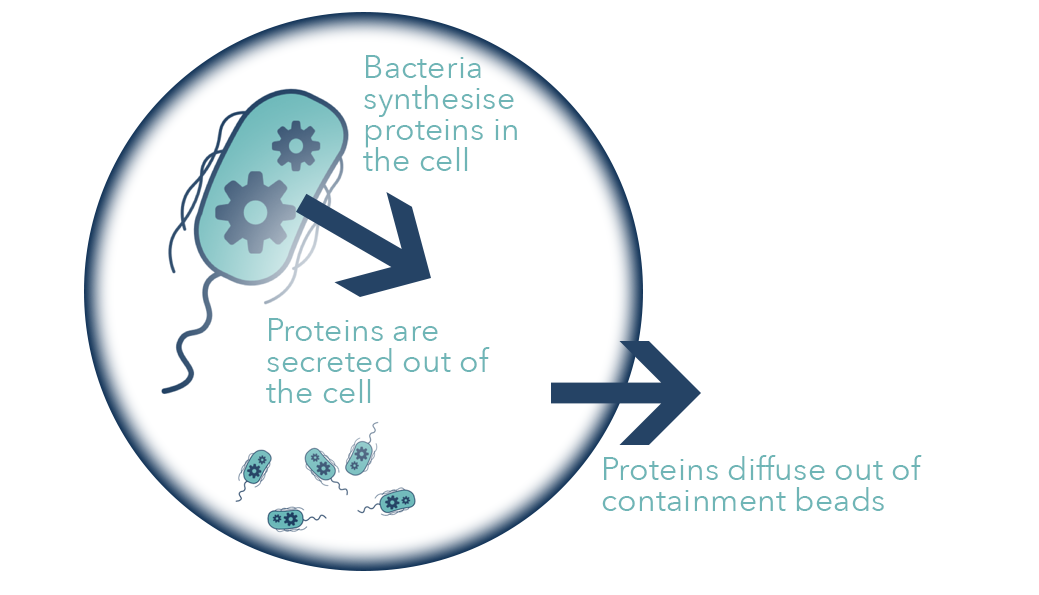
These beads could also be contained outside the body inside a bag of sterile water; this bag would then be plugged into a 3-way Foley catheter. This solution would then be washed through the catheter and into the bladder, therefore tackling any infection that may be present in the lining of the bladder.

First attempt at making the beads using Sodium Algniate
To see our process of designing and making the beads, look here.
Proof of Principle
Chemistry in Making the Beads
The gel created to encapsulate the bacteria is made of Calcium Alginate; this is synthesised when aqueous Sodium Alginate solution is dropped into Calcium Chloride solution.
Calcium Alginate is a water-insoluble gelatinous polymer; therefore it forms beads when the sodium ions are exchanged with the calcium ions. Each calcium ion can bond with two alginate polymer chains; this is called cross-linking. As the sodium ions can only bind to one polymer this cross-linking doesn’t occur and the polymer is water soluble, so the gel does not form.

Tutorial Video
Experimental Data
Firstly, we wanted to show that we could get the bacteria inside of the beads. To do this we created beads that contained fluorescent bacteria. The bacteria we used were from the interlab study: 20K MG, 20K ∆F, and 20K DH5, with MG(-) as a negative control.

We made sets of the beads for 5 days while measuring the fluorescence using the GFP protocol on the FLUOstar Omega plate reader.
The data shows that the wells that contain beads made with encapsulated fluorescent bacteria have a much higher fluorescence than the beads made without any bacteria and the beads made with the negative control bacteria. Therefore, we concluded that the bacteria are encapsulated inside of the beads.
We then made beads using Crystal Violet dye in order to record the diffusion of the dye out of the bead. The data could then be related to the diffusion of the protein via dimensional analysis for our modelling.
We collected the following spectrophotometric absorption data which we calibrated to prepared concentration standards.

(Further analysis of this can found in our modeling section.)
Next we tried to determine whether the bacteria were alive inside the beads and could therefore grow and continue to secrete our proteins.
To do this we made up beads containing the 4 different types of bacteria and placed these bacteria into wells of a 96-well plate containing M9 media. We measured the fluorescence of each well every 15 minutes over 20 hours at 37˚C.
This experiment produced very promising data that suggests the bacteria are growing inside the beads and producing GFP. Therefore we can assume that our bacteria would be able to grow and produce our anti-biofilm agents while encapsulated. The graph shows that the fluorescence of the 3 beads containing the fluorescent bacteria increased over time, and that the bead containing no bacteria remain constantly low. The bead containing our negative control had a very slight increase in fluorescence over time but much less than the bacteria containing the gene for GFP. This could be due to some contamination during the process of making the beads.

We put some consideration into the storage of our beads as hospitals would need to have a supply of them in order to carry out treatment effectively without having to make the beads and use them in a specific amount of time. This would cause delay in treatment and also a lot of waste of materials.
Our first idea was to freeze the beads, however when they are frozen the structure of the alginate is compromised and the skeleton of the beads begin to break down. Therefore, we looked at storing the beads at 4˚C and then investigated whether or not the bacteria would again secrete GFP after being warmed up to 37˚C.

From this we obtained some promising data. Beads stored in the cold room for 20 days were taken out and warmed to 37˚C. Then we measured the fluorescence of these beads every 15 minutes for 20 hours. The graph of this data shows that fluorescence does increase over time, indicating that the bacteria are growing inside of the beads and continuing to produce GFP.
An initial issue with our beads was that the beads had to be kept immersed in media in order to ensure a supply of nutrients to the encapsulated bacteria. To get around this, we tried making beads with our media as the solvent so the beads would have media encapsulated inside them, and thus the bacteria could still survive if the beads were placed into water/urine/etcetera. This internal nutrient supply would help to increase the longevity of the bacteria over the duration of the treatment, with the result that the catheter would have to be replaced less frequently - thus reducing the discomfort and risk of infection for the patient.
The beads were made up using 30mL of LB + Chl and 0.36g of sodium alginate. We then placed the resulting beads into the plate reader and measured the fluorescence of these beads every 15 minutes for 20 hours. We plotted the data on the graph below:

The graph demonstrates that the bacteria do grow inside of the beads, but they reach stationary phase much quicker than the beads what were surrounded by media. This could be due to the lower concentration of nutrients available.

We then took a bead and looked at it underneath a confocal microscope in order see into the bead. This gave us some incredible images of the bacteria fluorescing within the beads.
This photo shows clearly that GFP-producing bacteria are encapsulated inside the bead. This is our main evidence that we can get bacteria to survive in the beads.
Using confocal microscopy, we also took images of multiple layers of a bead and stacked them together to build up a 3D image of the bead and the bacteria encased within.

We then began to probe the edge of the bead with the miscroscope to see whether an bacteria had begun to leak out. From the image below we can see that there is a definition between the edge of the bead, where inside you can see the bacteria fluorescing and outside you cannot.

The Next Steps
If we had longer to work on our beads there are several things we could attempt in order to improve their viability.
The first would be to try and coat the beads and provide a second physical barrier to prevent the escape of the bacteria. We had the idea of coating them in a second layer of the calcium alginate, or to have another chemical in the Calcium Chloride bath that would instantly form a coating around the bead as it formed. However, this would require much more complex chemistry and material science - beyond what we could attempt in 10 weeks.
Ideally we would also make the beads much smaller, giving the beads a smaller volume, and so more would be able to fit inside of the catheter – while simultaneously improving the rate of diffusion out of the beads due to the relative increase in surface area. This would require a needle with a smaller lumen. The approach of making the beads could easily be scaled up so they could be produced in bulk.

Semi-Permeable Membrane
We would use a selectively permeable membrane to contain the beads within a compartment inside the catheter. This membrane would also provide an additional level of physical defence against our bacteria escaping into the urinary tract. The membrane would be similar to that used in a dialysis machine, and thus would allow urine and protein to diffuse through while preventing the larger bacteria from passing. Our largest protein is approximately 40kDa, so the diameter of the membrane pores would have to be able to accommodate this, as well as the larger components of urine (in order to prevent damning and a build up behind the membrane, causing complications). We would most likely be using one of the many materials already used for dialysis tubing membranes: polysulfone, polyethersulfone, etched polycarbonate, or collagen.
After considering the various options, we decided that for our catheter polysulfone would be best as it is most the commonly used in the medical profession and in dialysis machines. It is also reasonably simple to control the size of the pores during synthesis and thus produce the desired pore size.

Coating
To prevent biofilm formation on the outside of the catheter we would immobilize bacteria in a gel that would coat the outer surface. One option would be to form the gel into fibres that could be wrapped around the catheter and fixed in place with medical adhesive. The advantage to this is that the fibres would have a large surface area to volume ratio, ensuring greater diffusion of our DNase and DspB out of the gel and improving the anti-biofilm effect. A similar approach has been taken with previous catheter designs that are currently on the market, with coatings that contain silver nanoparticles, antibiotics etcetera.
An alternative would be to encase our bacteria in a thin sol-gel film; keeping our bacteria confined to the catheter surface but still allowing them to maintain their activity from within the gel. The properties of the gel could be adjusted so that it would be hydrophilic, aiding the insertion of the catheter and improving the comfort for the patient while in place. Our kill-switch design would be particularly useful here as it could be used to kill and bacteria that escaped the gel matrix. Engineering the kill-switch would take too long when compared to the timescale of our project so is not something we have attempted; however there is published literature that has shown it to work.
Manufacturing
Currently catheters are very cheap to produce, and thus as part of our project we decided to look into how we could introduce the beads into the pipe during the manufacturing process. We spoke to a mechanical engineer, Steve Dinsdale, and he helped us with a design for a machine that could do this. The machine would use the technique of extrusion to make the tube and then a second cooled inflow tube would introduce the beads. This would hopefully keep the bacteria cool enough so they are not killed, while the polymer at a high enough temperature to remain thermoplastic and form the tube.

Manufacturing idea from mechanical engineer Steven Dinsdale
Potential problems:
- If this design were used for catheter production the bacteria could potentially not survive the sterilisation process at the end of the manufacturing.
- Steps further down in the manufacturing process could harm our bacteria.
- The bacteria may not survive in storage due to the length of time stored, temperature, etc