Difference between revisions of "Team:Dundee/Forensic Toolkit/Fingerprints"
| Line 87: | Line 87: | ||
<figure align="center"> | <figure align="center"> | ||
| − | <img class="report-img" src="https://static.igem.org/mediawiki/2015/ | + | <img class="report-img" src="https://static.igem.org/mediawiki/2015/9/90/LSpurificationIMACDundee2015.jpg"> |
<figcaption class="report-img"> | <figcaption class="report-img"> | ||
<p><b>Figure 5 -</b>Purification of lanosterol synthase by nickel IMAC. A) Chromatogram showing the purification profile of the His-tagged haptoglobin. The fractions labelled (A8-A12) were further analysed by SDS page gel. B) 10µl of the labelled fractions were mixed with 10µl laemmli buffer and loaded onto a 12.5% SDS-PAGE gel and stained with Coomassie Blue. </p> | <p><b>Figure 5 -</b>Purification of lanosterol synthase by nickel IMAC. A) Chromatogram showing the purification profile of the His-tagged haptoglobin. The fractions labelled (A8-A12) were further analysed by SDS page gel. B) 10µl of the labelled fractions were mixed with 10µl laemmli buffer and loaded onto a 12.5% SDS-PAGE gel and stained with Coomassie Blue. </p> | ||
| Line 96: | Line 96: | ||
<figure align="center"> | <figure align="center"> | ||
| − | <img class="report-img" src="https://static.igem.org/mediawiki/2015/ | + | <img class="report-img" src="https://static.igem.org/mediawiki/2015/9/90/LSpurificationIMACDundee2015.jpg"> |
<figcaption class="report-img"> | <figcaption class="report-img"> | ||
<p><b>Figure 6 -</b>Elutions corresponding to the peaks from the nickel IMAC were collected ran on a 12.5% SDS-PAGE gel and then transferred to a nitrocellulose membrane and probed with an anti-His antbody. The band detected in the blot corresponds to the thick banding detected in Fig 4, at 37kDa.</p> | <p><b>Figure 6 -</b>Elutions corresponding to the peaks from the nickel IMAC were collected ran on a 12.5% SDS-PAGE gel and then transferred to a nitrocellulose membrane and probed with an anti-His antbody. The band detected in the blot corresponds to the thick banding detected in Fig 4, at 37kDa.</p> | ||
Revision as of 01:48, 19 September 2015
Abstract
Lanosterol synthase (LSS) an oxidosqualene cyclase (OSC) enzyme that specifically binds to squalene epoxide (2,3- oxidosqualene), which is present in fingerprints. We successfully managed to clone lanosterol synthase into pSB1C3 and the over expression vector pQE80-L. We successfully characterised its production by a Western immunoblot using an anti-His antibody. We then continued on in the hope to purify this enzyme from E. coli using Nickel IMAC.
Introduction
Fingerprints are defined as the ridge impressions left on a surface by a fingertip. They are the most commonly used form of criminal evidence globally, either equating or outnumbering all other forms of forensic evidence combined. A typical fingerprint is composed of 95% water with the remaining 5% being a mixture of both organic and inorganic compounds from the eccrine sweat and sebaceous glands. Figure 1 illustrates the key variables that interact and influence the composition of a fingerprint.

Figure 1 - This figure illustrates the relationship between fingertip, substrate and environmental conditions which collectively form a fingerprint and its constituents.
Lanosterol synthase is an enzyme that converts squalene epoxide to lanosterol in the pathway from squalene to cholesterol (Fig 2).

Figure 2 -Components involved in the second step of the pathway from Squalene to Cholesterol. A. Squalene epoxide configuration, steroid downstream from squalene. B. Lanosterol synthase crystal structure, the enzyme that converts squalene epoxide to lanosterol. C. Lanosterol configuration, the steroid donstream fro squalene epoxide.
Fingerprints are used in forensics as visual identification due to the unique ridge patterns between individuals. In dermatoglyphics, the scientific study of fingerprints, there is a key distinction between the terms ‘fingerprint’ and ‘fingermark’. Fingerprints are defined as the intentional imprint on a surface for recording purposes and often with the use of enhancement reagents to make the impressions distinct and fluorescent. ‘Fingermarks’ are incomplete (either smudged or distorted) impressions deposited incidentally on surfaces. Latent fingermarks are the most common type of fingerprint evidence found at crime scenes. The often blurred ridges add an extra depth of complexity when trying to link the fingermark found at a crime scene to the fingerprint of a suspect. Despite the heavy usage of fingerprint evidence for criminal investigations, the forensic procedure comes with its limitations. The most common evasions of prosecution in court are:
- The fingerprints of the suspect were found on a movable object.
- There is an absence of evidence as to when the fingerprints were placed.
Both arguments could be effectively eliminated if a test existed that could date fingermarks left at crime scenes. The area of dating fingermarks based on their chemical composition is a relatively unexplored field in forensic sciences. This is due to the lack of reliability of proposed techniques. It has been suggested in theory, it may be possible to age fingerprints based on quantitative changes of kinetic compounds found in fingerprints.
Following the information obtained from the modelling team (link to mathematical modelling page), squalene was found to be the best compound to target within the fingerprint. Squalene was found to be present up to 21 days once the fingerprint was deposited. When talking to fingerprint experts it was suggested that it would be more beneficial to identify a component that could narrow down the age of a fingerprint to one week or less. Squalene is an intermediate in the pathway to the production of Cholesterol and looking down from squalene we identified squalene epoxide.

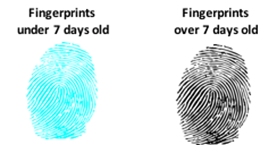
Figure 3 -Using nanobeads to target squalene epoxide (S.E) contained in the fingerprint to age the print. S.E is present up until the 7th day after deposition and therefore a fingerprint can be roughly aged. The concentration of S.E increases over the first 5 days as well leading to the possibility of narrowing it down more closely.
Using fluorescent microspheres, we would like to detect the presence of squalene epoxide through the use of its specific enzyme lanosterol synthase. By fusing lanosterol synthase to the fluorescent nanobead, when applied to a fingerprint the nanobead would bind to squalene epoxide, if present, and therefore fluoresce. The current procedure of detecting quantities of chemical compounds in fingerprints is gas chromatography and mass spectrometry (GC/MS). Despite the accuracy the current technique provides it comes with the flaw that it completely destroys the fingerprint evidence. A fluorescent biosensor device comes with the advantage of keeping the evidence intact for other analyses.
Results
Firstly, Human Lanosterol Synthase (LSS) was optimised for expression in E.coli and modified to ensure it was compatible with BioBrick specifications and standards. This gene was then synthesized by IDT (Integrated DNA Technologies ) and we then succesfully cloned it into pSB1C3 and submitted it as a biobrick (BBa_K1590006)
The synthetic gene was then sub-cloned into the pQE80-L overexpression vector that adds an N-terminal hexa-histidine tag onto the protein which allows for purification by immobilized metal affinity chromatography (IMAC).
Initial experiments were carried out to optimize expression of lanosterol synthase within our E.coli chassis. Fig 4 shows the expression of LSS within E.coli.

Figure 4 -Detection of His-tagged LSS in whole cells of E.coli. Single colonies of E.coli strain M15 pREP4 harbouring LSS. Cells were used to inoculate 5ml of LB growth medium supplemented with 100µg/ml ampicillin and 50µg/ml Kanamycin. Once the OD600 reached 0.7 the cells were then induced with IPTG, as indicated. Cells were then grown for a further 4 hours at 37°C, 1ml aliquots were pelleted and cells reuspended in 100µl laemmli buffer and 20µl of samples were separated by SDS-PAGE (12% acrylamide) and transferred to nitrocellulose membrane and probed with anti-His antibody.
As shown in fig 4, there is successful production of lanosterol synthase (expected mass 83kDa) along with traces of degradation. Upon successful expression of LSS- His, expression was then scaled up and 4 litre cultures were grown up for purification of lanosterol synthase by nickel IMAC. The results from this can be seen in Figure 5.

Figure 5 -Purification of lanosterol synthase by nickel IMAC. A) Chromatogram showing the purification profile of the His-tagged haptoglobin. The fractions labelled (A8-A12) were further analysed by SDS page gel. B) 10µl of the labelled fractions were mixed with 10µl laemmli buffer and loaded onto a 12.5% SDS-PAGE gel and stained with Coomassie Blue.
Lanosterol synthase was expected to be 83kDa, however the majority of protein purified is at 37kDa. This was further analysed by carrying out a western blot to detect for any full length LSS- His purified.

Figure 6 -Elutions corresponding to the peaks from the nickel IMAC were collected ran on a 12.5% SDS-PAGE gel and then transferred to a nitrocellulose membrane and probed with an anti-His antbody. The band detected in the blot corresponds to the thick banding detected in Fig 4, at 37kDa.
The results obtained from the blot showed no presence of lanosterol synthase at 83kDa, suggesting we are only detecting a degraded version of it instead of the whole enzyme. The purification conditions will have to be optimized in order to continue with our fingerprint aging device.
Future Work
After purification and characterisation, through site-specific mutagenesis, the active sites (positions 232 and 455) will have to be inactivated to guarantee that the enzyme wouldn’t destroy any vital evidence on the scene or convert the present squalene epoxide into lanosterol.

Figure 7 -Crystal structure of lanosterol synthase with the active sites at positions 232 and 455 (red). The yellow arrows indicate the active sites in laonsterol synthase that will have to be mutated to inactivate the enzyme to avoid unwanted activity.
References
- Jones, B. Comprehensive Medical Terminology: A Competency-Based Approach. 3rd ed. Thomson Delmar Learning, USA; 2008.
- Adebisi, S. S. Fingerprint Studies-The Recent Challenges and Advancements: A Literary View. The Internet Journal of Biological Anthropology, 2, 1-15; 2009.
- Llewellyn, P. Jr., Dinkins, L. New use for an old friend. J. Forensic Identif. 1995; 42:498–503.
- Yamashita, B., French, M. in: J. Barnes (Ed.) Fingerprint Sourcebook, NCJ 225320. US Department of Justice, Washington; 2011
- Frick, Amanda Akiko. Chemical investigations into the lipid fraction of latent fingermark residue; 2015.


