Team:Cambridge-JIC/MicroMaps
Image Stitching
This is the technology that makes MicroMaps all possible...and the reason it is in early alpha. Though the translation mechanism of our microscope allows panning control as fine as a single micron (see our Tech Specs page), the material used for 3D-printing and the quality of the motors used impairs the accuracy with which translation can be achieved. A subtle point of the flexure mechanism, used for stage movement, is that shifting in one direction, also causes a small angle twist of the frame of view. This in turn makes it difficult to know which parts of the slide go where in our interface. Luckily, image stitching algorithms have been created to find where two or more images match and combine them together (such as in the panorama feature on modern smartphones). Using these algorithms we can determine precisely how the microscope imagery should be shown on screen and eliminate the seams between them. We can also use the position information derived to correct for translational inaccuracies so we can know with confidence where you are on your slide and enable you to drop pins on features you like. In addition, the overlaying of the stitched images actually removes the black artefacts deposited by dirt on the CCD or imperfections of the optics.
There are many stitching algorithms that have been developed, though unfortunately some of the best are proprietary. As an open-source alternative, our algorithm is significantly less robust and this has been a major roadblock in the development of MicroMaps. Eventually, we were forced to disable the free panning mechanism on MicroMaps alpha. It has also meant that some of other features, that we had in mind, have not been integrated for now.
Algorithms required for MicroMaps:
feature detecting algorithms: (these find interesting things in images)
proprietary: SIFT, SURF, FAST
free: ORB - actually this algorithm is also pretty good, according to its developers [2]
feature MATCHING algorithms: (these try to match the same interesting things in two images)
proprietary: FLANN - fast and reliable
free: BF - Brute-Force, unreliable
Our research in the area indicates that the SIFT + FLANN combination is very good. Further understanding of the subject might be gained from Google's PhotoSphere project.
Examples:

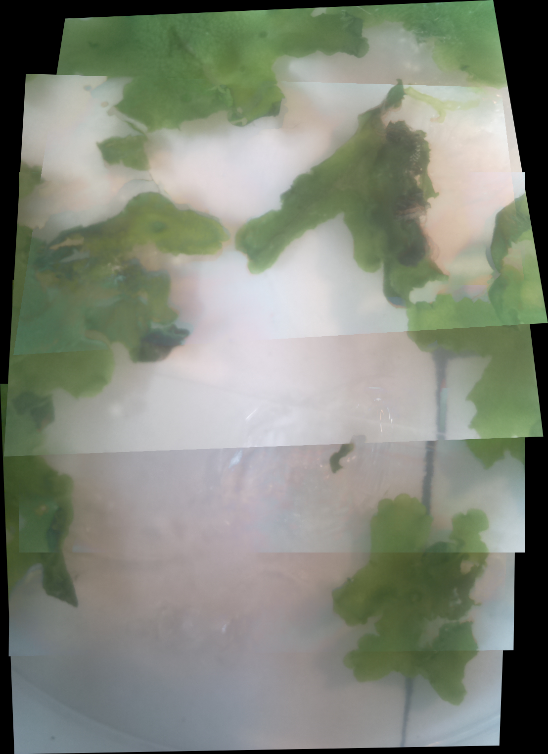

Figure 1: First successful stitching of two images (Nigerian liane). Figure 2: Stitching implemented on macroscopic images of Marchantia polymorpha as part of our WebShell. We are still working on this issue, expect the potential fix in MicroMaps Version 2. Made some progress with an image tracking challenge: each of our software team was challenged to track some moving pixels in an image ***(ants.gif) to find the hidden message, Will's entry shown in bants.gif***. Perhaps in V.2 interesting live features (eg. moving cells) will be automatically tracked.


