Team:Exeter/Interlab

Interlab Study
Prologue: The Devices
The aim of the interlab study was to measure the strength of three constitutive Anderson promoters, using GFP as a measure of strength, in order to generate data which can then be compared between different iGEM labs. Here we tell our story of the interlab study, including the promoters which were tested, and the ways in which we measured their strength. Follow the chapters in order using the buttons above to be guided through this story. We begin in this section by stating the promoters which were measured, and the devices these were assembled into in order to measure them, along with the controls used.
Devices 1, 2 and 3:
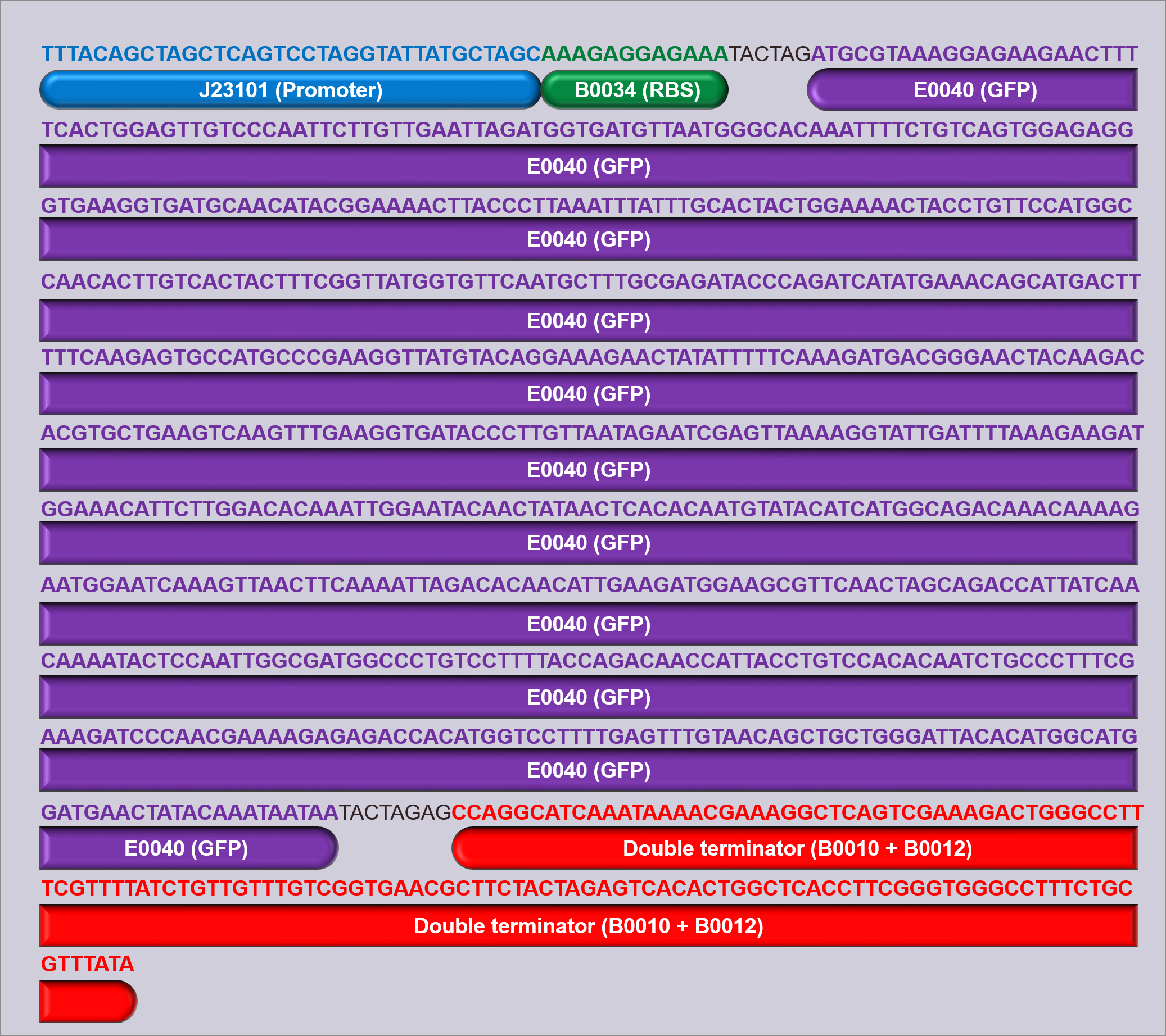
Three devices were constructed in order to test the promoters; D1, D2 and D3. Each device contains a promoter, followed by the GFP measurement composite part I13504 (figure 1). This part contains the GFP biobrick BBa_E0040, which is preceded by a strong RBS (B0034) and succeeded by a double terminator (B0010 and B0012). By using this same part in all of the devices constructed, it is ensured that the only variation between devices is the promoter, and the use of a strong RBS ensures that the expression of GFP should be limited by the strength of the promoter, allowing measurement of promoter strength.
The promoters used in devices 1, 2 and 3 were J23101 (D1), J23106 (D2), and J23117 (D3) respectively (figure 1), and all devices were built into and expressed in a pSB1C3 plasmid backbone. The pSB1C3 plasmid confers chloramphenicol resistance, which was used to select for cells containing the device. Once built, all of the devices were sequenced verified (figure 2, also available for download here).
The Controls
In the measurement of the above devices, three controls were used; a GFP positive control (I20270), a GFP negative control (R0040), and a cell growth control (untransformed cells). I20270 contains the same RBS, GFP gene, and double terminator, but contains the J23151 promoter. J23151 is also a constitutive promoter, allowing for a direct comparison to the promoters in the devices built, as well as acting as a positive GFP control. R0040 contains a Tet repressible promoter, meaning that it will not fluoresce and can act as a GFP negative control during measurement. Figure 3 shows schematics of these parts.
Chapter 1: New Beginnings
Once upon a time, there was an iGEM team from the University of Exeter. Determined to contribute to the field of synthetic biology, we decided to participate in the Second International Interlab Measurement Study. While determining fluorescence levels for the three Interlab devices, we hoped to gain some experience in the lab before starting our iGEM project, Ribonostics. We aimed to build and measure the fluorescence of three BioBrick devices:
- J23101 + I13504 (D1)
- J23106 + I13504 (D2)
- J23117 + I13504 (D3)
In the first week of our iGEM induction, we started out by hydrating DNA from the kit, after which we followed the standard protocols for BioBrick assembly. The first step was transforming our BioBrick DNA into competent DH5α cells (New England Biolabs), and growing them in overnight cultures. After transforming, we performed our first miniprep to obtain the DNA needed for further experiments, and checked the DNA concentration using Qubit. We then used values obtained from this to calculate the volumes of all of components of the digestion step. After the digestion, we ran a part of each sample on a gel to see if the insert and vector were the correct size. The ligation was the last step; after this, we plated our samples and incubated them overnight. We hoped that the next morning, there would be glowing green colonies on our plates. Protocols of these experiments can be found on these pages of the lab book: [transformation, miniprep, Qubit, digestion, ligation]
The next day, we came into the lab, hoping to see colonies so green they would be blinding. After viewing the plates under blue light to visualise the green fluorescent protein (GFP) in our constructs, we understood that the statement ‘biology sometimes doesn’t work’ is entirely accurate. There were no glowing colonies on our plates. However, this moment of truth inspired us to diagnose what was wrong with our devices, and give the Interlab Study another try in the following week.
Our lab induction and Interlab Study were a good chance to find out where the equipment and resources in the lab were, as well as to meet some of the researchers working there – many of them helped at various stages of our project. Those amongst us with little or no lab experience learned basic techniques, such as pouring agar and making LB broth, but also made an effort to understand the biological concepts behind the Interlab Study. One of our physicists, Todd, said that in the first week, he learned the skills necessary for any budding biologist – pipetting, making, and pouring agar. ‘I thought it was very useful. It was great.’ – a direct quote from Todd. Equipped with these skills, we set out to complete the Interlab Study accurately, efficiently, and most importantly – successfully.
Chapter 2: Under Pressure
After a weekend of thought, we came into the lab, determined to get glowing colonies by the end of the week. The answer to why our Interlab Study failed the previous week was simple: we had been trying to put the wrong BioBricks into the pSB1C3 plasmids – they did not join, because they could not join.
We looked at our second attempt at the Interlab Study with optimism – we had some practice in the lab, and we understood more clearly what it was we were actually trying to do. This time, we made and used TOP10 competent cells. Again, we followed the same flowchart of experiments, using the correct BioBricks (J23101, J23106, J23117).
This time, only three or four of us worked in the lab at any one time. During our first week in the lab, we learned that having more people in the lab does not necessarily mean we are more productive. During the second try at the Interlab Study, we also made sure we had all the necessary controls – our competent TOP10 cells alone, the empty pSB1C3 backbone, a working GFP plasmid and a negative control of the TetR plasmid. After a few setbacks (such as forgetting to put LB broth during the incubation stage of a bacterial transformation), we arrived at yet another overnight incubation, and again, we asked ourselves the burning question – would we see glowing colonies?
Chapter 3: We Fight On
By now, you may have guessed that our team is not the luckiest when it comes to glowing. If so, you guessed right – we did not have any glowing colonies.
We were baffled, puzzled, surprised – but not broken. Once again, we marched into the lab, determined to see fluorescent colonies. We double (and triple) checked that we were using the correct BioBricks, and enzymes during the digestion step. We also thought carefully about what we could change in order to get our Interlab Study to work. During our third attempt, we followed a protocol for a traditional digestion instead of the digestion protocol we had used before. We also altered the transformation protocol slightly. After running our digest on the gel, we performed a gel extraction – this way we would be sure that we were using vector and insert fragments of the correct sizes. Amongst the things we added and changed were also: control plates of just our competent cells, and using 1 µl of freshT4 ligase instead of 0.5 µl. Again, we followed the Interlab flowchart of procedures. Armed with our transformed Interlab samples and plenty of controls, we saw no reason for our colonies not to glow the next morning.
Chapter 4: New Direction
On the rainy Friday morning, we climbed the stairs to the fourth floor, where our lab was located. We had great – but realistic – expectations for our colonies. Once we saw them, we knew the likely result of our third attempt at the Interlab Study. The blue light confirmed our suspicions – we did not have any green glowing colonies. We had a discussion about what could be going wrong with the Interlab process, and came up with three hypotheses:
- During our ligation, we used a 1:1 ratio of insert to vector – perhaps using a 3:1 or 1:3 ratio would be better.
- We always incubated our ligation overnight in the fridge – maybe incubating at room temperature would work more efficiently.
- Our antibiotic plates had been in the cold room for a long time – had the antibiotic somehow broken down?
Nevertheless, the problem was the ligation step of the Interlab procedures. We considered our options, and decided that we would continue with the Interlab Study, but in a different way: we would construct our three devices using IDT gBlocks, assemble them using the Gibson method, transform into E. coli cells and measure their fluorescence. There were a number of reasons why we chose to do this: time was the main issue. Designing the constructs for Ribonostics took up the majority of our time, and our team was becoming disjointed, with some members focusing entirely on the Interlab Study. The primary purpose of the Interlab Study is to measure the strength of three promoters in different labs, and we figured that we would be able to fulfil this goal with our new method. We became even more determined to complete the Interlab Study, and re-evaluated our goals:
- After assembling our three devices, we would measure them using the TECAN microplate reader in our lab.
- After this, we would perform FACS on our devices to measure fluorescence of each individual cell.
- We would subsequently use ImageStream to take pictures of our cells.
- Expressing the Interlab Study constructs cell-free would be another one of our goals: this would allow us to compare fluorescence in cells and cell-free. Full of hope and determination, we ordered the gBlocks that would later be assembled into our three devices.
Chapter 5: Seeing Red
.It took a few weeks for the gBlocks to arrive in our lab. We used this time to prepare for our next attempt at the Interlab study – plan our experiments, find and understand the protocols, especially ones relating to gBlock assembly, which none of us had done before. Once the sequences arrived, a dedicated team of three – Bradley, Jasmine and Georgina – went to the lab to attack the Interlab Study again. gBlocks were prepped and constructed according to the protocols, transformed into DH5α cells, then plated and incubated overnight. Our standard positive and negative controls were used, as well as the NEB positive control from the gBlock assembly kit.
Life is full of surprises, as our team found out the next morning. For the first time, we saw glowing colonies – but they were not glowing green. They were red. We figured out that the plasmid in which our Interlab sequences were in, pSB1C3, which is made from RFP parts, had reformed.
The first glowing colonies we obtained during the Interlab Study – the next step will be to get them to glow the right colour.In the afternoon, two of us went back to the lab, and picked five non-red colonies and one red colony from the plates for each device, as well as one colony from a positive control. We were hoping to see some green glowing after growing them out overnight. However, the colonies we got back were limited in number and did not glow. Looking for an explanation, we decided to miniprep samples from each device, and sequence them to find out exactly what had happened.
So once again the fluorescence promised to us by our interlab constructs had eluded us again, since we knew the wait for our sequencing results could take a while, we decided to move on and start with the gBlocks from scratch. However half way through this process we received the results back from our first gBlock sequences and it had worked! This left us with more questions, if the gBlocks had worked then why were they not glowing? So we proceeded to use a different machine to measure fluorescence, picking TECAN which showed clearly that they did indeed fluoresce just not as brightly as we had expected. With this in mind we started to plan all the things we could measure.


