Difference between revisions of "Team:Exeter/Results"
| Line 224: | Line 224: | ||
<h2>Further characterisation:</h2> | <h2>Further characterisation:</h2> | ||
| + | <p> | ||
| + | The following parts were further characterised: | ||
| + | <ul> | ||
| + | <li><a href="http://parts.igem.org/Part:BBa_K1073020">aeBlue</a></li> | ||
| + | <li><a href="http://parts.igem.org/Part:BBa_K1073022">eforRed</a></li> | ||
| + | <li><a href="http://parts.igem.org/Part:BBa_K1431814">amajLime</a></li> | ||
| + | </ul> | ||
| + | </p> | ||
<h4>Absorbance spectra:</h4> | <h4>Absorbance spectra:</h4> | ||
| + | |||
| + | |||
| + | <div class="containerSlider300" style="float:left"> | ||
| + | <div class="slider300"> | ||
| + | |||
| + | <div class="slide"><a href="https://static.igem.org/mediawiki/2015/4/4b/Exeter_aeBlue_spectrum.png"><img src="https://static.igem.org/mediawiki/2015/4/4b/Exeter_aeBlue_spectrum.png" title="Figure 4: Spectrum for aeBlue, peak maxima at 595nm."></a></div> | ||
| + | <div class="slide"><a href="https://static.igem.org/mediawiki/2015/4/40/EforRed_spectrum.png"><img src="https://static.igem.org/mediawiki/2015/4/40/EforRed_spectrum.png" title="Figure 4: Spectrum for eforRed, peak maxima at 580nm."></a></div> | ||
| + | <div class="slide"><a href="https://static.igem.org/mediawiki/2015/4/4b/Exeter_aeBlue_spectrum.png"><img src="https://static.igem.org/mediawiki/2015/4/4b/Exeter_aeBlue_spectrum.png" title="Figure 4: Spectrum for amajLime, peak maxima at 595nm."></a></div> | ||
| + | |||
| + | </div> | ||
| + | </div> | ||
| + | |||
| + | |||
| + | <p> | ||
| + | Shown in figure 4 are the absorbance spectra for aeBlue, eforRed, and amajLime between 400nm and 850nm. The peak maxima for these are 595nm, 580nm, and 595nm respectively. | ||
| + | </p> | ||
| Line 232: | Line 256: | ||
| + | <div class="containerSlider300" style="float:left"> | ||
| + | <div class="slider300"> | ||
| + | <div class="slide"><a href="https://static.igem.org/mediawiki/2015/8/86/Exeter_amajLime_standard_curve.png"><img src="https://static.igem.org/mediawiki/2015/8/86/Exeter_amajLime_standard_curve.png" title="Figure 5: Standard curve for amajLime measured at 458nm."></a></div> | ||
| + | <div class="slide"><a href="https://static.igem.org/mediawiki/2015/a/a0/Exeter_aeBlue_standard_curve.png"><img src="https://static.igem.org/mediawiki/2015/a/a0/Exeter_aeBlue_standard_curve.png" title="Figure 5: Standard curve for aeBlue measured at 595nm"></a></div> | ||
| + | <div class="slide"><a href="https://static.igem.org/mediawiki/2015/d/dc/Exeter_eforRed_standard_curve.png"><img src="https://static.igem.org/mediawiki/2015/d/dc/Exeter_eforRed_standard_curve.png" title="Figure 5: Standard curve for eforRed measured at "></a></div> | ||
</div> | </div> | ||
| + | </div> | ||
| + | |||
| + | <p> | ||
| + | Figure 5 shows standard curves of concentration in mg ml<sup>-1</sup> vs. OD at peak maxima for each of the three chromoproteins further characterised. | ||
| + | </p> | ||
| + | |||
| + | </div> | ||
| + | |||
| + | <h4>Standard curve - concentration vs. OD:</h4> | ||
Revision as of 02:01, 19 September 2015
Results
Toehold experiments
In silico testing:
Experimental validation of GreenFET1J (BBa_K1586000):
As was explained on the experiments page, GreenJ's (BBa_K1586000) function was characterised and validated by measuring fluorescence intensity of GFP in response to increasing amounts of trigger RNA. Shown in figure 1 is the raw data which was collected from this experiment. In order to analyse this data, the log10 of the amounts of TrigGreen (in nanograms) were calculated. So that the log10 value for 0ng could be calculated, 1x10⁻⁶ng was used instead, and all values were increased by six so that 0ng shows as 0 on the graph. These data were then plotted on a graph and a linear trend-line fitted (figure 2).
Further characterisation:
The following parts were further characterised:
Absorbance spectra:
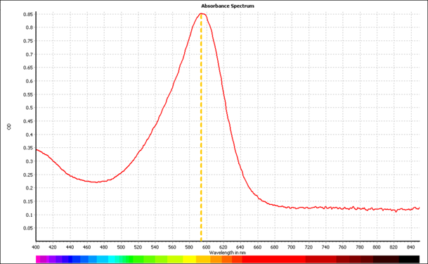
Shown in figure 4 are the absorbance spectra for aeBlue, eforRed, and amajLime between 400nm and 850nm. The peak maxima for these are 595nm, 580nm, and 595nm respectively.
Standard curve - concentration vs. OD:
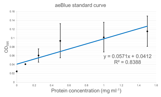
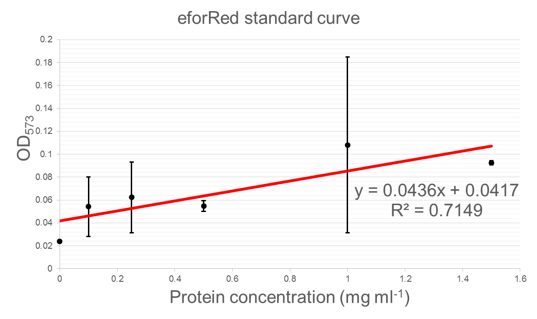
Figure 5 shows standard curves of concentration in mg ml-1 vs. OD at peak maxima for each of the three chromoproteins further characterised.


