Difference between revisions of "Team:Cambridge-JIC/Description"
Maoenglish (Talk | contribs) |
KaterinaMN (Talk | contribs) |
||
| (85 intermediate revisions by 3 users not shown) | |||
| Line 1: | Line 1: | ||
{{:Team:Cambridge-JIC/Templates/Menu}} | {{:Team:Cambridge-JIC/Templates/Menu}} | ||
<html> | <html> | ||
| + | |||
| + | <style> | ||
| + | |||
| + | .dark:hover | ||
| + | { | ||
| + | color:#78a87f; | ||
| + | } | ||
| + | |||
| + | hr | ||
| + | { | ||
| + | border-color:#78a87f; | ||
| + | background-color:#78a87f; | ||
| + | color:#78a87f; | ||
| + | } | ||
| + | </style> | ||
| + | |||
| + | <style> | ||
| + | section.guide-section a:link, section.guide-section a:visited{ | ||
| + | color:#6fd85e !important; | ||
| + | } | ||
| + | img | ||
| + | { | ||
| + | filter: grayscale(1); | ||
| + | -webkit-filter: grayscale(1); | ||
| + | -moz-filter: grayscale(1); | ||
| + | -o-filter: grayscale(1); | ||
| + | -ms-filter: grayscale(1); | ||
| + | } | ||
| + | |||
| + | img:hover | ||
| + | { | ||
| + | filter: grayscale(0); | ||
| + | -webkit-filter: grayscale(0); | ||
| + | -moz-filter: grayscale(0); | ||
| + | -o-filter: grayscale(0); | ||
| + | -ms-filter: grayscale(0); | ||
| + | } | ||
| + | } | ||
| + | </style> | ||
| + | |||
| + | |||
| + | <section style="background-color: #fff"> | ||
| + | <div class="slide" style="position:relative"> | ||
| + | <div style="width: 100%; padding: 0% 10%; margin: 30px 0px;color:#000;position:absolute;top:50%"> | ||
| + | <center><p><span class="dark" style="font-size:340%">Microscopy awaits you...</span></p></center> | ||
| + | </div></div></section> | ||
| + | |||
| + | <section style="background-color: #3d3d3d"> | ||
| + | <div class="slide" style="position:relative"> | ||
| + | <div style="width: 100%; padding: 0% 10%; margin: 30px 0px;color:#fff;position:absolute;top:50%"> | ||
| + | <center><p><span class="dark" style="font-size:170%">Cambridge-JIC brings you OpenScope: the foundation to a new era of accessible microscopy.</span></p></center> | ||
| + | </div></div></section> | ||
| + | |||
<section style="background-color: #fff"> | <section style="background-color: #fff"> | ||
<div class="slide"> | <div class="slide"> | ||
| − | <div style="width: | + | <div style="width: 100%; padding: 0% 10%; margin: 30px 0px;color:#000"> |
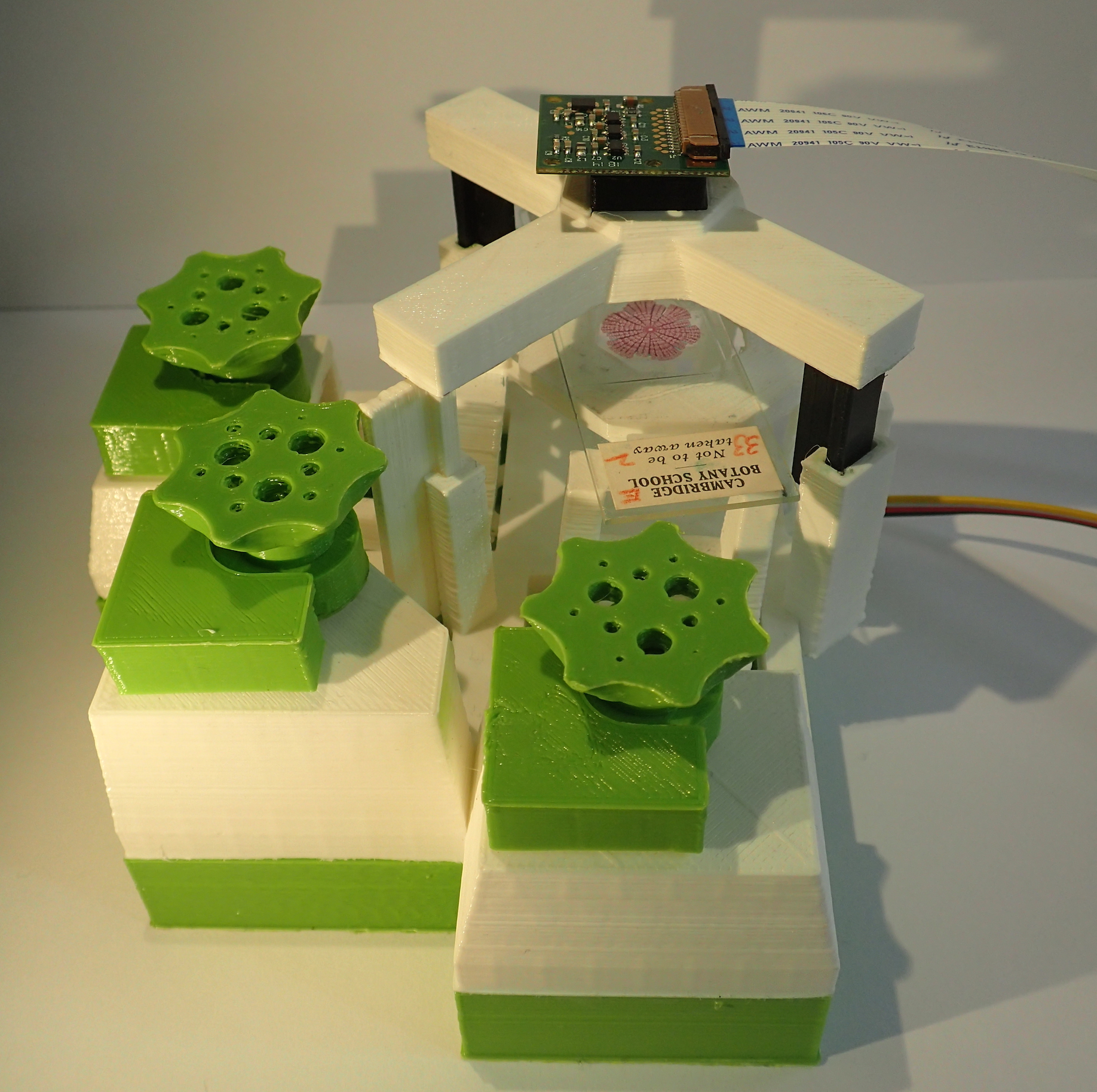
| − | + | <p><span class="dark" style="font-size:170%">The chassis</span> is 3D printed, allowing simple modification. The plastic is cheap, biodegradable and flexible. Stage translation, based on work by Dr Richard Bowman [1], makes use of the flexibility to give fine control.</p> | |
| − | < | + | <hr> |
| − | + | <p><span class="dark" style="font-size:170%">The mechanics</span> of the stage can be automated using stepper motors. The user has remote control of the microscope, and can introduce tailor-made programs to facilitate their experiments.</p> | |
| + | <hr> | ||
| + | <p><span class="dark" style="font-size:170%">The optics</span> are low-cost, low-energy and modular. Illumination using LEDs means reducing power consumption and cost. A Raspberry Pi camera makes the microscope digital, and an epi-fluorescence cube makes imaging GFP a reality. With sub-micrometer resolution in brightfield and darkfield modes, you are ready to image single cells or whole tissues.</p> | ||
| + | <hr> | ||
| + | <p><span class="dark" style="font-size:170%">The software</span> uses OpenCV and forms a core part of the project. The Webshell gives you real-time control over the microscope live-stream: from time-lapse to scale-bars. MicroMaps uses image stitching and sample recognition algorithms to give you the whole sample field in one, ready for annotation and screening. Autofocus capabilities allow automation of OpenScope’s motors, letting you image dynamic samples without supervision.</p> | ||
| + | <hr> | ||
| + | <p><span class="dark" style="font-size:170%">The documentation</span> is comprehensive, non-proprietary and easy to access. And its licensed to make sure it stays that way.</p> | ||
| + | <hr> | ||
| + | <p><span class="dark" style="font-size:170%">The community</span> of ‘makers’ is free to develop, modify and redistribute the documentation. OpenScope can evolve, improve and adapt to different needs.</p> | ||
| + | <hr> | ||
| + | <p><span class="dark" style="font-size:170%">The overall cost</span> is below £200, orders of magnitude below commercial lab microscopes.</p> | ||
| + | <p style="font-size:80%">[1] Sharkey, J., Foo, D., Kabla, A., Baumberg, J. and Bowman, R. (2015). <i>A one-piece 3D printed microscope and flexure translation stage.</i><a href="http://arxiv.org/abs/1509.05394" class="blue">[online]</a> Arxiv.org. [Accessed 18 Sep. 2015].</p> | ||
| + | </div></div></section> | ||
| − | + | ||
| − | < | + | |
| − | + | <section style="background-color: #3d3d3d"> | |
| − | < | + | <div class="slide"> |
| − | < | + | <div style="width: 100%; padding: 0% 10%; margin: 30px 0px;color:#fff"> |
| − | < | + | <p><span class="dark" style="font-size:170%">The result?</span></p> |
| − | < | + | <hr> |
| − | <p style="font-size: | + | <p>A microscopy solution for synthetic biologists, letting <span class="dark" style="font-size:170%">researchers</span> tailor the microscope to their needs. Image in the incubator, fume-hood or the field using remote access and battery power, or use OpenScope for rapid preliminary screening.</p> |
| − | + | <hr> | |
| − | < | + | <p>A microscopy solution for <span class="dark" style="font-size:170%">schools</span>, providing education in programming, optics and one of the most ubiquitous techniques in biological research: fluorescence imaging.</p> |
| − | + | <hr> | |
| − | </p> | + | <p>A microscopy solution for laboratories with small budgets, based on low-cost and easily sourced components. The potential of Openscope to increase access to microscopy in <span class="dark" style="font-size:170%">developing countries</span> has been a key part of the design process from day one. </p> |
| − | --> | + | |
</div> | </div> | ||
</div> | </div> | ||
</section> | </section> | ||
| + | |||
| + | |||
| + | <section style="background-color: #fff"> | ||
| + | <div class="slide" style="position:relative"> | ||
| + | <div style="width: 100%; padding: 0% 10%; margin: 30px 0px;color:#000;top:20%;position:absolute"> | ||
| + | <center> | ||
| + | <img src="//2015.igem.org/wiki/images/e/e4/CamJIC-gallery7.jpeg" style="height:240px;margin:10px"> | ||
| + | <img src="https://static.igem.org/mediawiki/2015/6/6f/CamJIC-OpenScope_Everything.JPG" style="height:240px;margin:10px"> | ||
| + | <img src="https://static.igem.org/mediawiki/2015/f/fd/CamJIC-OpenScope_Above.jpeg" style="height:240px;margin:10px"> | ||
| + | |||
| + | </center> | ||
| + | |||
| + | </div></div></section> | ||
</html> | </html> | ||
{{:Team:Cambridge-JIC/Templates/Footer}} | {{:Team:Cambridge-JIC/Templates/Footer}} | ||
Latest revision as of 21:17, 18 September 2015